Warning: mkdir(): Permission denied in /home/virtual/lib/view_data.php on line 86 Warning: fopen(/home/virtual/tipjournal/journal/upload/ip_log/ip_log_2024-04.txt): failed to open stream: No such file or directory in /home/virtual/lib/view_data.php on line 88 Warning: fwrite() expects parameter 1 to be resource, boolean given in /home/virtual/lib/view_data.php on line 89 Deep Sulcus Sign Developed in Patient with Multiple Fibrous Bands between the Parietal and Visceral Pleura
TIP Search

CLOSE


Trauma Image and Procedure > Volume 2(1); 2017 > Article
Park, Yeo, Park, Kim, Lee, Kim, Cho, and Yeom: Deep Sulcus Sign Developed in Patient with Multiple Fibrous Bands between the Parietal and Visceral Pleura

Abstract

A deepening of the costophrenic angle occurs in cases with a deep sulcus sign. We report a case of deep sulcus sign in a 47-year-old man who fell from the fifth floor. Supine chest radiography showed a right-sided pneumothorax with deep sulcus sign. Chest computed tomography (CT) demonstrated a large pneumothorax with multiple fibrous bands between the parietal and visceral pleura of the upper lobe of the right lung.

CASE

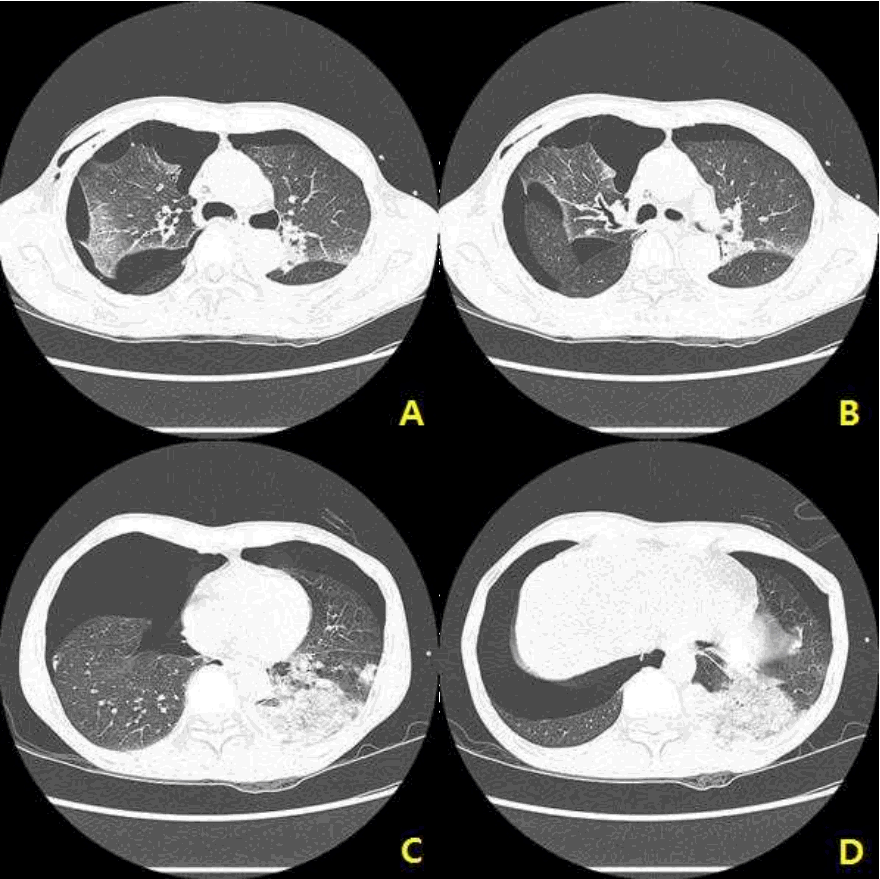
A 47-year-old man presented to the emergency department after falling from a fifth floor height. His vital signs were systolic blood pressure 60 mmHg, pulse rate 111 beats/min, respiration rate 31 breaths/min, body temperature, 36.4℃, and oxygen saturation 96%. The injury severity score was 29, revised trauma score 5.15, trauma and injury severity score 74.8%. His arterial blood gas analysis was pH 7.35, pCO2 29 mmHg, pO2 75 mmHg, hemoglobin 16.7, SaO2 94%, lactic acid 11.8 mmol/L, and base excess -8.0. Supine chest radiography showed a right-sided pneumothorax with a deep sulcus sign (Fig. 1.). The axial views of chest computed tomography (CT) demonstrated a large pneumothorax with multiple fibrous bands between the parietal and visceral pleura of the upper lobe of the right lung (Fig. 2.). The coronal view of chest CT more clearly showed the deep sulcus sign seen in the chest AP (Fig. 3.). The deep sulcus sign disappeared after tube thoracostomy (Fig. 4.).

DISCUSSION

The deep sulcus sign on a supine chest radiograph is an indication of a pneumothorax [1]. The costophrenic angle is abnormally deepened when the pleural air collects laterally, producing the deep sulcus sign [1]. Gordon [1] first reported a deep sulcus sign as a sign of pneumothorax in 1980. Then, the deep sulcus sign was reported on the right side in a trauma patient [2-4]; it is rarely reported on the left side [5]. A deep sulcus sign also was reported in nontraumatic patients [6], and Wang et al. [7] reported a deep sulcus sign caused by free air in the abdominal cavity.

Notes

Conflict of Interest Statement

No potential conflict of interest relevant to this article was reported.

Fig. 1.
Supine chest radiograph shows a right-sided pneumothorax with a deep sulcus sign.
tip-2-1-7f1.gif
Fig. 2.
Chest CT (coronal view) demonstrates a large pneumothorax with multiple fibrous bands between the parietal and visceral pleura of the upper lobe of the right lung.
tip-2-1-7f2.gif
Fig. 3.
Chest CT (coronal view) more clearly shows the deep sulcus sign.
tip-2-1-7f3.gif
Fig. 4.
Supine chest X-ray after the tube thoracostomy shows the disappearing of the deep sulcus sign.
tip-2-1-7f4.gif

REFERENCES

1. Gordon R. The deep sulcus sign. Radiology. 1980;136(1):25-7.
crossref pmid
2. Kim HH, Park CY, Cho HM. Deep Sulcus Sign. Trauma Image and Procedure. 2016;1(1):12-3.
crossref pdf
3. Liu S-Y, Tsai I-T, Yang P-J. Pneumothorax and deep sulcus sign. QJM: An International Journal of Medicine. 2016;109(9):621-2.
crossref
4. Sabbar S, Nilles EJ. Deep sulcus sign. New England Journal of Medicine. 2012;366(6):552 -.
crossref pmid
5. Smally AJ, Suozzi JC. Images in emergency medicine. Deep sulcus sign. Annals of emergency medicine. 2007;49(5):717 , 25.
crossref pmid
6. Tseng YH, Liu KL, Shih IL. Deep sulcus sign. Emergency medicine journal : EMJ. 2012;29(7):608.
crossref pmid
7. Wang SH, Wei KY, Liu YC, Huang CH. The deep sulcus sign indicates free air in the abdomen. Internal and emergency medicine. 2015;10(8):1033-4.
crossref pmid
TOOLS
Share :
Facebook Twitter Linked In Google+
METRICS Graph View
  • 0 Crossref
  •    
  • 8,256 View
  • 46 Download


ABOUT
ARTICLE CATEGORY
Article Category

Browse all articles >

BROWSE ARTICLES
FOR AUTHORS AND REVIEWERS
Editorial Office
Korean Association for Research, Procedures and Education on Trauma
#1618, 18, Mapo-daero 4da-gil, Mapo-gu, Seoul 04177, Korea
Tel: +82-2-3280-9013    E-mail: karpet@karpet.or.kr

Copyright © 2024 by Korean Association for Research Procedures and Education on Trauma.

Developed in M2PI

Close layer
prev next