Endovascular Repair of Traumatic Common Iliac Vein Rupture
Article information
Abstract
Iliac vein rupture after blunt trauma is extremely rare, but there are some case reports describing endovascular repair of this lesion. However, there is no case report describing insertion of a stent graft in a patient with return of spontaneous circulation (ROSC) after cardiac arrest. Here we report our experience of performing stent graft insertion on a patient with ROSC with left common iliac vein rupture.
CASE
A 64-year-old male patient was transferred to our emergency department. He had experienced a return of spontaneous circulation (ROSC) after cardiopulmonary resuscitation (CPR) (performed owing to cardiac arrest) and was transferred to our hospital. On the abdominal computed tomography (CT) at the original hospital, a contrast leak was observed in the left common iliac vein. After arriving at our hospital, mechanical ventilation was initiated, and cardiac arrest and ROSC occurred twice more. After a massive transfusion, his blood pressure was 96/50 mmHg, and radiologic intervention was performed. Venography, which was done through puncture of the left common femoral vein, revealed rupture and massive extravasation (Fig. 2.). Through a 12F sheath, a 16–12 mm/100 mm stent graft (Gore Excluder stent graft) was inserted. Subsequent venography revealed disappearance of the extravasation (Fig. 3.). After the intervention, the patient’s blood pressure recovered to 124/70 mmHg, and he underwent no further treatment in the intensive care unit. However, the status of the patient deteriorated, and he died the day after the intervention.
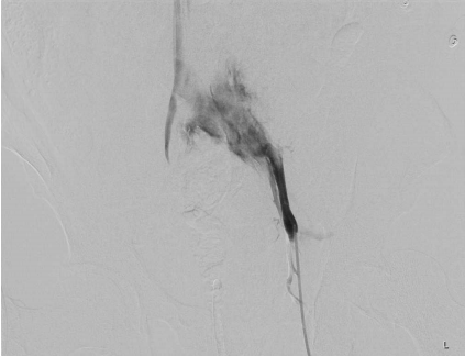
Venography using left femoral access shows massive extravasation from the left common iliac vein.

Post–stent-graft venography shows disappearance of the contrast extravasation from the left common iliac vein.
DISCUSSION
Blunt trauma-induced iliac vein rupture is rarely reported [1-3]. Until now, there has been no case report describing stent insertion in an iliac vein in a patient with ROSC after CPR. Endovascular management of iliac vein rupture can be an option to consider for a patient whose hemodynamic status is relatively stable.

Axial view of a computed tomography scan of the portal venous phase shows a left iliac bone fracture and contrast extravasation from the left common iliac vein.
Notes
CONFLICT OF INTEREST
No potential conflict of interest relevant to this article was reported.