


Introduction
Central venous catheterization is a common procedure performed in the emergency department and intensive care unit; thereafter sufficient training is required for adequate catheter placement. Among various insertion routes, most subclavian vein access may be placed using anatomic landmarks alone [1]. This study primarily aimed to present the central venous catheterization procedure via the subclavian vein.
Main body
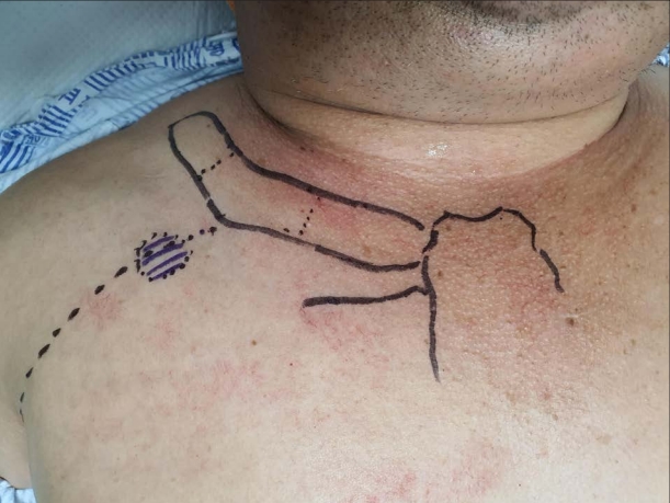
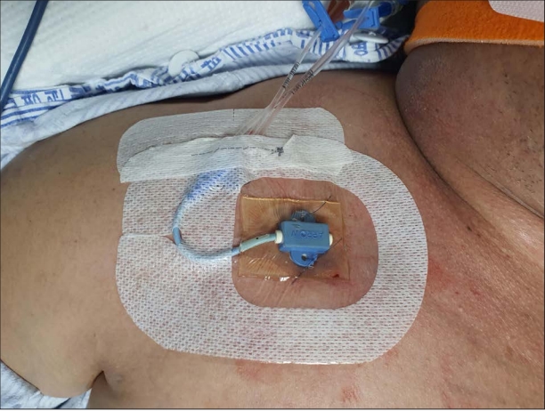
The subclavian vein continues to the axillary vein, which runs from the lateral border of the first rib to the medial border of the anterior scalene muscle, just below the clavicle and toward the sternal notch. At preprocedure, the patient is positioned in the Trendelenburg position, which increases venous dilation and reduces the risk of air embolus [1]. To facilitate initial access, a shoulder roll may be placed along the patient’s upper thoracic spine [2]; however, the shoulder roll is typically omitted when a spinal injury is suspected. Next, the access site is determined. While the right side had a lower incidence of pneumothorax, it had a higher incidence of catheter malposition. After taking maximal sterile barrier precautions, such as wearing a head cap, face mask, sterile body gown, and gloves [3], skin preparation with chlorhexidine solution is advised for the upper region of the chest wall, from the top of the shoulders to the nipple line, and the patient’s neck and chin [4]. After a full-size sterile drape has been placed, only the prepared area is exposed. All three lumens at the 7-Fr triple lumen ARROWg+ ard Blue® CVC (Arrow, Zdar, Czech Republic) should be flushed with sterile saline before inserting the needle. Typically, the access point is two fingers caudal and lateral to the clavicle angle, at approximately two-thirds of the way lateral from the sternal notch (Fig. 1). After injecting local anesthesia into the skin, the operator positions one hand on external landmarks, placing the index finger in the sternal notch and the thumb in the clavicle angle, whereas the other hand controls the puncture needle. The puncture needle is then advanced through the skin at the access point to the clavicular angle. After the puncture needle touches the clavicle, it is slightly retracted and advanced parallel to the clavicle’s line toward the sternal notch. A steep angle should be avoided between the needle and clavicle to prevent pneumothorax. While attempting venous cannulation, a syringe is placed on mild suction. A burst of dark-red blood confirms access to the subclavian vein, and this should flow in a nonpulsatile pattern after withdrawing the syringe. The pulsatile pattern identifies an arterial puncture, although it may be difficult to elicit the typical flow in patients with shock. Once the arterial puncture has been confirmed, the needle should be removed and the region compressed. Moreover, any return of air into the syringe barrel should be cautiously monitored, which indicates a pleural space break and the possibility of pneumothorax [5]. After confirming that the guidewire is in the vein, the guidewire is advanced through the needle into the vein. If any resistance is encountered, the guidewire should be withdrawn. A syringe should be used to confirm that the needle is in the vessel by observing regurgitation, and then, the guidewire should be reinserted. Some recommendations were indicated for positioning the guidewire’s tip appropriately. First, the needle is turned 90° clockwise before inserting the guidewire. The needle tip is sloped so that a 90° clockwise rotation helps the guidewire from entering the internal jugular vein. Second, the main body of guidewire faces the patient’s body. The guidewire is coiled in a circular main body so that its natural course can aid in achieving the right position. Third, the patient’s neck is bent toward the insertion site and pulled toward the chin, if possible. This helps prevent the guidewire from entering the internal jugular vein by compressing it. When a guidewire tip contacts the right atrium, arrhythmias might occur. By slightly withdrawing the guidewire, these arrhythmias can be easily controlled. After successfully placing the guidewire, the needle is removed. Guidewire control should be maintained at all times so that it does not entirely enter the vein. A tiny skin incision with the blade is made at the access point that is sufficiently large to allow the dilator and catheter to pass without resistance. The dilator is then passed across the guidewire with maintaining the angle between the skin and the guidewire while keeping at least one hand on the wire at all times. Some mild resistance is typically observed as the dilator passes through the subcutaneous soft tissues. The dilator is then withdrawn from the guidewire, followed by catheter placement over the guidewire. This procedure is performed with the insertion site compressed with the left middle fingertip. When the guidewire appears through the brown port of the catheter, the end of the guidewire is held by the opposite hand, and the catheter is advanced over the guidewire. The ideal catheter tip placement around the cavoatrial junction is generally 15 cm from the right access point and 18 cm from the left access point [6]. After positioning a catheter with the appropriate length, the guidewire is removed, and all ports are aspirated and flushed to ensure access. The white rubber grip and blue plastic clamp are applied around the catheter next to the insertion site and securely attached to the patient’s skin with 3-0 nylon sutures. The catheter hub is also sutured. A sterile dressing should be placed after completing the procedure (Fig. 2). A postprocedure chest X-ray should be routinely performed to assess the catheter tip location and possible complications such as pneumothorax.
All procedures were recorded at the trauma center of Dankook University Hospital (Video 1).