Warning: mkdir(): Permission denied in /home/virtual/lib/view_data.php on line 86 Warning: fopen(/home/virtual/tipjournal/journal/upload/ip_log/ip_log_2024-04.txt): failed to open stream: No such file or directory in /home/virtual/lib/view_data.php on line 88 Warning: fwrite() expects parameter 1 to be resource, boolean given in /home/virtual/lib/view_data.php on line 89 Migration of intracranial air according to position change in post-traumatic pneumocephalus: erect and supine skull X-ray views
TIP Search

CLOSE


Trauma Image and Procedure > Volume 5(1); 2020 > Article
Park, Ahn, Kang, Jung, Seo, and Moon: Migration of intracranial air according to position change in post-traumatic pneumocephalus: erect and supine skull X-ray views

Abstract

Pneumocephalus is often detected in patients with trauma. However, it is difficult to find studies on position change of intracranial air according to the patient position observed on skull X-rays (erect and supine views). This is because skull X-rays are rarely performed in erect position for patients with head injury. We report a case of location change of intracranial air associated with patient position change observed in the skull X-ray of a patient with traumatic pneumocephalus. To the best of our knowledge, no such cases have previously been reported in English literature.

Introduction

Traumatic pneumocephalus is caused by air collection in the intracranial cavity after trauma. When intracranial air causes intracranial hypertension, it leads to tension pneumocephalus, which results in mass effect with neurological deterioration [1-3]. Pneumocephalus is generally diagnosed on brain computed tomography (CT) but is rarely identified on skull X-ray, as observed in this case [4].

Case presentation

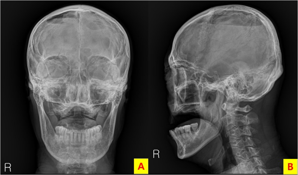
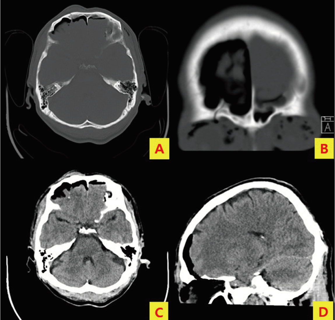
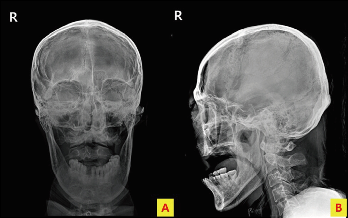
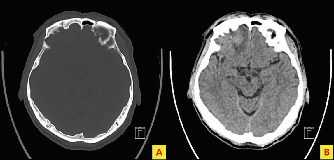
A 59-year-old man was referred to our emergency department from another hospital after falling forward from a ladder, on which he was working, at a height of 1.5 m. He complained of facial pain, had facial swelling, and his Glasgow Coma Scale was 15. Initial vital signs were as follows: blood pressure of 176/88 mmHg, pulse rate of 87 beats/min, respiration rate of 16 breaths/min, body temperature of 37.4°C, and oxygen saturation of 94%. Skull X-ray (anteroposterior [AP] and lateral views) performed in erect position at the previous hospital showed intracranial air in the parietal region (Fig. 1), and fracture of the frontal bone and pneumocephalus were observed on brain CT (Fig. 2). Skull X-ray (AP and lateral views) in supine position and brain CT performed at our hospital showed intracranial air in the frontal region (Fig. 3). Conservative treatment was adjusted, and brain CT performed on the 10th day of admission showed that the intracranial air had nearly disappeared (Fig. 4). The patient was discharged on the 14th day of admission without any complications .

Discussion

Traumatic pneumocephalus rarely progresses to tension pneumocephalus, which is accompanied by neurological deterioration and should be considered a neurosurgical emergency [1-3]. Pneumocephalus is primarily diagnosed on brain CT and is rarely observed on skull X-ray [4]. In this case, skull X-ray was performed in the erect position at another hospital, which showed presence of intracranial air in the parietal region. In contrast, at our hospital, skull X-ray was performed in the supine position, which showed presence of intracranial air in the frontal region. Observing intracranial air migration caused by position change in skull X-rays is extremely difficult because skull X-ray in erect position is rarely performed in case of patients with head injury. To the best of our knowledge, no such case has been reported in English literature. In our case, there was no neurological deterioration, conservative treatment was administered, and subsequent brain CT revealed gradual disappearance of the intracranial air.

Notes

Conflict of interest

No potential conflict of interest relevant to this article was reported.

Fig. 1.
Skull X-ray (anteroposterior [AP] and lateral views) performed in erect position at a local hospital showing intracranial air in the parietal region.
tip-5-1-13f1.jpg
Fig. 2.
Brain computed tomography (CT) performed at a local hospital showing fracture and subdural pneumocephalus.
tip-5-1-13f2.jpg
Fig. 3.
Skull X-ray (anteroposterior [AP] and lateral views) performed in supine position at our hospital showing intracranial air in the frontal region.
tip-5-1-13f3.jpg
Fig. 4.
Brain computed tomography (CT) performed on the 10th day of admission showing nearly complete disappearance of the intracranial air.
tip-5-1-13f4.jpg

References

1. Al-Aieb A, Peralta R, Ellabib M, El-Menyar A, AlThani H. Traumatic tension pneumocephalus: Two case reports. Int J Surg Case Rep. 2017;31:145-9.
crossref pmid pmc
2. Clement AR, Palaniappan D, Panigrahi RK. Tension Pneumocephalus. Anesthesiology. 2017;127(4):710.
crossref pmid
3. Rao V, Fredriksli O, Gulati S. Post-traumatic epidural tension penumocephalus: a case report. J Med Case Rep. 2015;26(9):151.

4. Odebode OT, Afolabi OA, Suleiman OA. Lateral skull radiograph in a patient with posttraumatic tension pneumocephalus complicated by late epilepsy. Emerg Med J. 2008;25(1):32.
crossref
TOOLS
Share :
Facebook Twitter Linked In Google+
METRICS Graph View
  • 0 Crossref
  •    
  • 6,281 View
  • 32 Download


ABOUT
ARTICLE CATEGORY
Article Category

Browse all articles >

BROWSE ARTICLES
FOR AUTHORS AND REVIEWERS
Editorial Office
Korean Association for Research, Procedures and Education on Trauma
#1618, 18, Mapo-daero 4da-gil, Mapo-gu, Seoul 04177, Korea
Tel: +82-2-3280-9013    E-mail: karpet@karpet.or.kr

Copyright © 2024 by Korean Association for Research Procedures and Education on Trauma.

Developed in M2PI

Close layer
prev next