Usefulness of C-Arm Fluoroscopy in Treating Patients Injured by a Shotgun
Article information
Abstract
Shotgun injury, a rare event, is difficult to treat. Moreover, it is annoying and difficult to locate and remove bullets from patients using surgery. Here, we describe here a shotgun injury case wherein we removed the bullets using C-Arm fluoroscopy.
CASE
A 79-year-old male with no medical history was admitted to the emergency room with multiple shotgun injuries. He was mistakenly shot by a hunter hunting for a water deer. Upon admission, the patient was hemodynamically stable, but showed multiple shotgun injuries on the head, chest, abdomen, forearm, and leg (Fig. 1.). Further, he had hemoperitoneum and peritonitis because of bowel perforation and a mesentery injury. We accordingly planned for an emergency abdominal surgery followed by an orthopedic surgery (Fig. 2.). We employed C-Arm fluoroscopy to accurately locate the bullets in his body and to check for any remaining bullets. All bullets were removed using C-Arm fluoroscopy (Fig. 3.). The patient recovered without any complications.
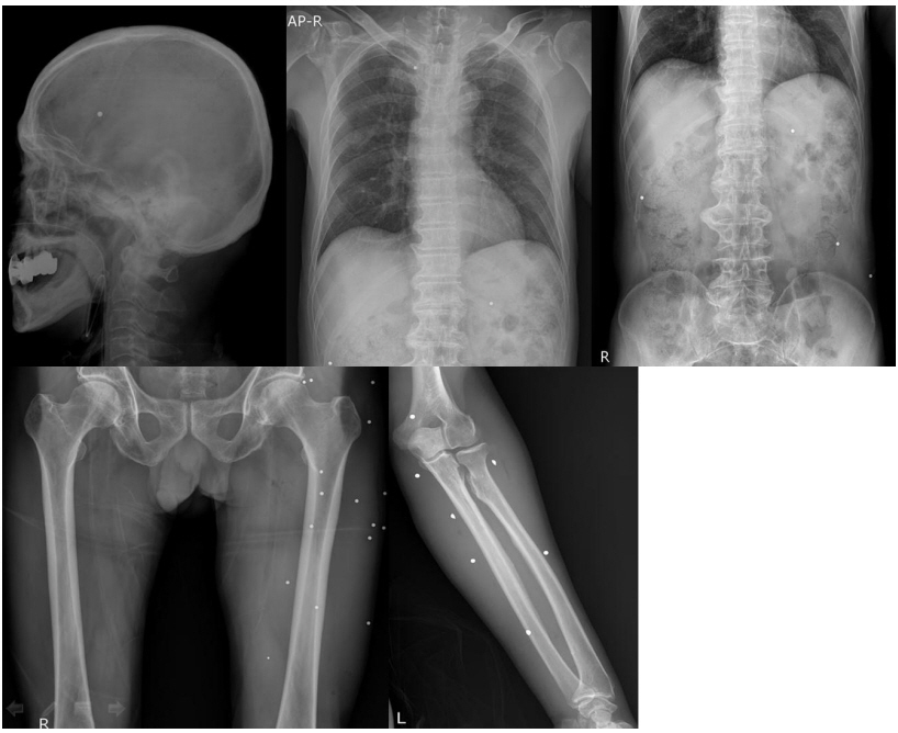
Plain X-Ray indicating bullet wounds at multiple sites
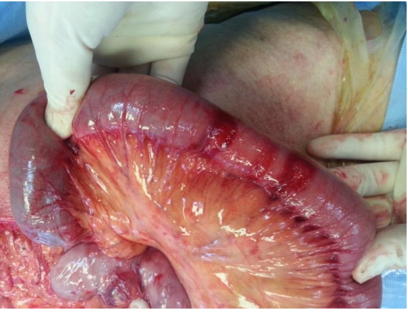
Surgical finding of a small bowel injury
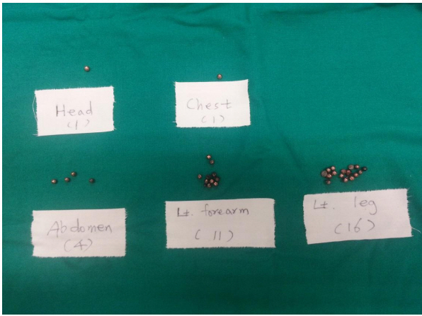
Total bullets removed from the patient
DISCUSSION
High-velocity firearm bullets travel at a speed of >2000 ft/s and transfer high energy to the tissues, whereas those of low-velocity weapons travel at <1000 ft/s. Shotgun is an example of the latter type. A shotgun cartridge contains pellets of varying sizes (very small to large) [1]. Therefore, it is difficult to locate and remove these bullets from patients injured by a shotgun. In conclusion, C-Arm fluoroscopy is a useful modality for treating patients injured by a shotgun.
Notes
Conflict of Interest Statement
No potential conflict of interest relevant to this article was reported.