Warning: mkdir(): Permission denied in /home/virtual/lib/view_data.php on line 93 Warning: chmod() expects exactly 2 parameters, 3 given in /home/virtual/lib/view_data.php on line 94 Warning: fopen(/home/virtual/tipjournal/journal/upload/ip_log/ip_log_2024-09.txt): failed to open stream: No such file or directory in /home/virtual/lib/view_data.php on line 100 Warning: fwrite() expects parameter 1 to be resource, boolean given in /home/virtual/lib/view_data.php on line 101 Deep Sulcus Sign
TIP Search

CLOSE


Trauma Image and Procedure > Volume 1(1); 2016 > Article
Kim, Park, and Cho: Deep Sulcus Sign

Abstract

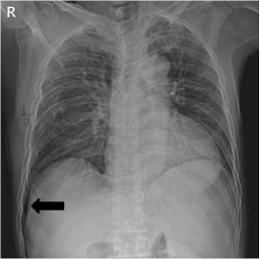
The deep sulcus sign is a radiolucent lateral sulcus where the chest wall meets the diaphragm. In a supine film, it may be the only indication of a pneumothorax because air collects anteriorly and basally within the nondependent portions of the pleural space, as opposed to the apex when the patient is upright. The costophrenic angle is abnormally deepened when the pleural air collects laterally, producing the deep sulcus sign. We present a brief image of a trauma showing the deep sulcus sign indicating a pneumothorax.

CASE

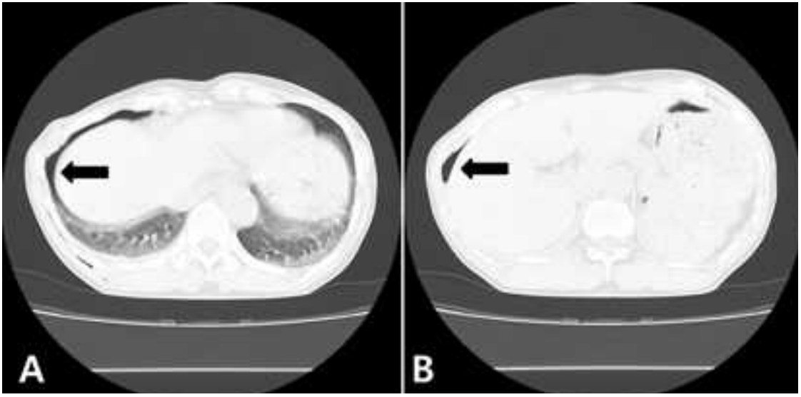
A 56-year-old male pedestrian was brought to the emergency department after a high-speed road-traffic accident. He had transient hypotension and tachycardia, which improved after the administration of intravenous fluids. The physical examination revealed multiple orthopedic injuries in addition to trauma to the right chest, pelvis, and head. He underwent prompt intubation and sedation. Chest radiography with the patient in the supine position showed a deep sulcus sign (Fig. 1.), which was highly suggestive of a pneumothorax. Chest computed tomography confirmed a pneumothorax (Fig. 2.).

DISCUSSION

In the supine position, air in the pleural space distributes anteriorly and basally at nondependent portions and causes deepening of the lateral costophrenic angle, producing the deep sulcus sign [1-3]. The deep sulcus sign is an important clue for diagnosing pneumothorax.

Notes

CONFLICT OF INTEREST

No potential conflict of interest relevant to this article was reported.

Fig. 1.
Chest anteroposterior view radiograph shows a right pneumothorax with deep sulcus sign.
tip-1-1-12f1.gif
Fig. 2.
Chest computed tomography revealed a pneumothorax.
tip-1-1-12f2.gif

REFERENCES

1. Jeng MJ, Lee YS, Tsao PC, Soong WJ. Neonatal air leak syndrome and the role of high-frequency ventilation in its prevention. Journal of the Chinese Medical Association : JCMA. 2012;75(11):551-9.
crossref pmid
2. Sabbar S, Nilles EJ. Images in clinical medicine. Deep sulcus sign. The New England journal of medicine. 2012;366(6):552.
crossref pmid
3. Wang SH, Wei KY, Liu YC, Huang CH. The deep sulcus sign indicates free air in the abdomen. Internal and emergency medicine. 2015;10(8):1033-4.
crossref pmid
TOOLS
Share :
Facebook Twitter Linked In Google+
METRICS Graph View
  • 1 Crossref
  •    
  • 18,214 View
  • 70 Download


ABOUT
ARTICLE CATEGORY
Article Category

Browse all articles >

BROWSE ARTICLES
FOR AUTHORS AND REVIEWERS
Editorial Office
Korean Association for Research, Procedures and Education on Trauma
#1618, 18, Mapo-daero 4da-gil, Mapo-gu, Seoul 04177, Korea
Tel: +82-2-3280-9013    E-mail: karpet@karpet.or.kr

Copyright © 2024 by Korean Association for Research Procedures and Education on Trauma.

Developed in M2PI

Close layer
prev next