Vertical Pelvic Fracture with Hemodynamical Stability
Article information
Abstract
Vertically unstable fractures of the pelvic ring, mostly caused by falls from heights or traffic accidents, are high-energy injuries with high mortality and morbidity rate. Patients with unstable pelvic ring fracture usually have hemodynamic instability, presenting a higher ratio of patients in shock already in the field. We described a case in which initial conservative treatment including fluid resuscitation and pelvic traction for vertical pelvic fracture with stable hemodynamics after a blunt trauma.
CASE
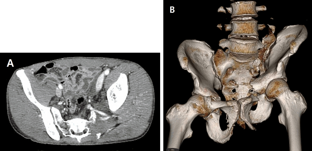
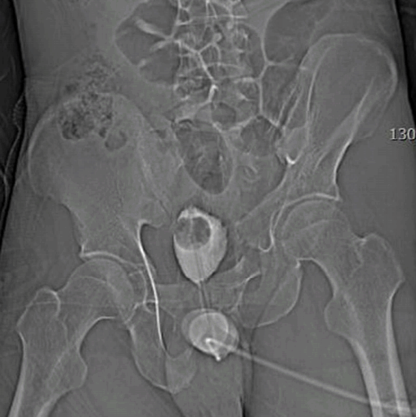
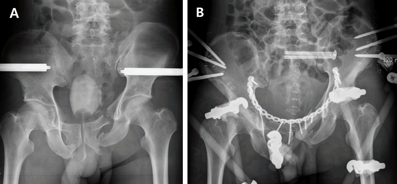
A 46-year-old man was ran over by an excavator and admitted to the emergency department. Upon arrival, he was alert with transient hypotension having a response to the transfusion of 2 units of packed red blood cells. The injury severity score was 29. The Focused Assessment with Sonography for Trauma showed no intraabdominal fluid collection. Supine pelvic X-ray showed multiple fractures in left pelvic with a dislocation (Fig. 1). Abdomen computed tomography scan revealed comminuted fractures and a vertical dislocation of left sacral ala and left pubic ramus with diastatic widening of left sacroiliac joint and hematoma around pelvic fractures without extravasation (Fig. 2). His stable hemodynamics was preserved without invasive procedures including angiographic embolization and massive transfusion. External pelvic fixation with C-clamp was performed the next day after skeletal traction applied to the left pelvicbone and lower extremity on admission day (Fig. 3A). Then, closed reduction with internal fixation of pelvis and anterior external pelvic fixation was performed in hospital day 7 (Fig. 3B).

(A) Abdomen computed tomography scan shows retroperitoneal hematoma of pelvis around left sacroiliac joint without extravasation, and (B) 3-D reconstruction of pelvis shows comminuted fractures and vertical dislocation of left sacroiliac joint, and bilateral fractures of pubic superior and inferior ramus.
DISCUSSION
The injury pattern of vertical pelvic fracture was rotationally and vertically unstable, with a complete disruption of the posterior osseous ligamentous structure of the pelvis [1]. Patients with unstable pelvic ring fracture, Young-Burgess classification types B and C showed significantly worse vital signs compared with type A, presenting a higher ratio of patients in shock already in the field for them [2]. This case was an unstable pelvic ring fracture, Young-Burgess classification type C, but hemorrhagic shock did not develop, not based on its injury severity. Initial resuscitative management was performed without massive transfusion, and operative management was carried out next day during the skeletal traction. Therefore, resuscitative treatment for pelvic fracture should be determined based on the hemodynamics and bleeding tendency of patient, with even unstable pelvic ring fracture.
Notes
Conflict of Interest Statement
No potential conflict of interest relevant to this article was reported.

