A Right-sided“J”-shaped Incision Provides Full Exposure of a Penetrating Wound from the Left Anterior Neck to the Right Thoracic Cavity: A Case Report
Article information
Abstract
We present a case report of a penetrating neck injury from the anterior neck to the right thoracic cavity. When we encounter such a case in the operating room, it is difficult to decide on an approach that exposes the entirety of the wound. In this case, only a partial sternotomy with a right anterior thoracotomy (extended “J”-shaped incision) provides full exposure of the injured area. Such an incision may be very effective in selective cases.
CASE
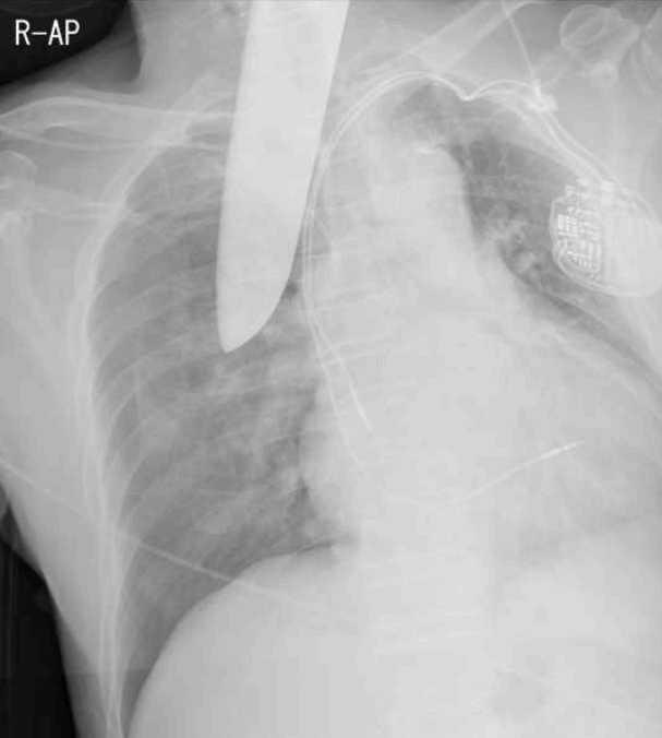
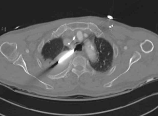
A 77-year-old man presented to the trauma bay with a self-inflicted neck stab wound (Fig. 1). Initial measurements of his vital signs revealed a blood pressure of 187/110 mmHg, pulse rate of 59 beats/min, respiration rate of 22 breaths/min, body temperature of 36.0°C, and oxygen saturation of 97%. He was drunk, and we could not check his mental state. The patient had a previously installed pacemaker because of a left bundle branch block. Initial chest X-rays showed that a knife traversed from the anterior region of the neck to the right thoracic cavity and induced a right pneumothorax (Fig. 2). A chest computed tomography (CT) scan showed a left lower neck laceration, thyroid gland injury, a foreign body in the mediastinum and the right hilum, and the tip was a posterior arc of the 6th rib (Fig. 3). We could not rule out injuries to the thyroid gland, trachea, and brachiocephalic and subclavian arteries and thus elected to perform an emergency operation. We performed a partial sternotomy and then extended to the right anterior thoracotomy. This incision looked like an extended “J” shape. With this incision, we could see the full injury area and were thus able to remove the knife and repair the lung, pleura, and thyroid and ligate small mediastinal vessels (Fig. 4). The patient was discharged on postoperative day 7 without any complication (Fig. 5).

Simple chest X-ray shows a right pneumothorax and a knife from the left anterior region of the neck to the right thoracic cavity.

Chest CT shows a foreign body in the left thoracic cavity and a right hemothorax and a laceration of the right-upper region of the lung.
DISCUSSION
Penetrating neck injuries occur in approximately 5–10% of all trauma patients who present to the emergency room. About 30% of these neck injuries are accompanied by trauma outside the neck zones as observed in the present case [1,2,3]. In the presence of right-sided lung and vascular injuries, the common surgical approach typically involves median sternotomy. With this incision, sometimes it is difficult to inspect the full injuries of the vessels and thoracic organs; therefore, a sternotomy with an extension of the anterior thoracotomy may be more useful in such situations. However, it is also known that a combination of an anterolateral thoracotomy, partial sternotomy, and left infra- or supraclavicular incision described as "trap-door" thoracotomy is rarely performed due to the time requirements and because it results in multiple fractures [4]. In the present case, the injury encompassed regions from left cervical zone 1 to the right thoracic cavity and the vital signs were stable. First, we planned to make a “trap-door” incision because subclavian vessel injuries could not be ruled out; however, after division of the manubrium and 3rd intercostal space, there were no subclavian vessel injuries. Thus, we were able to remove the knife, repair the lacerated lung, ligate the transected branches of the subclavian vessels, and repair the thyroid through this incision. During trauma surgery, it is challenging to expose arch vessels, their branches, and mediastinal structures; therefore, the selection of the incision depends on mediastinal structures that need to be explored during surgery [5,6]. The “trap-door” incision, or a modified version of this method, is an option in such cases.
Notes
Conflict of Interest
This work was supported by clinical research grant from Pusan National University Hospital in 2016.


