Developmental Dislocation of Hip Misdiagnosed as Traumatic Posterior Hip Dislocation
Article information
Abstract
A 47-year-old woman was presented to the emergency department. The limb of the patient was shortened, and her hip was mildly flexed, adducted, and internally rotated. Initially, the patient was misdiagnosed with a posterior hip dislocation. However, after careful history taking and radiologic evaluation, her final diagnosis was developmental hip dysplasia, which was a sequelae of poliomyelitis.
CASE
A 47-year-old woman was presented to our emergency department via another hospital. She fell from a 5-m height on a mountain. Immediate orotracheal intubation was performed owing to decreased mentality caused by acute subdural hematoma and subarachnoid hemorrhage. The medical records from the previous hospital noted failed manual reduction of the left hip. The external appearance of the patient’s left lower limb was shortened, and the hip was mildly flexed, adducted, and internally rotated. The patient’s family reported that she had a limping gait since early childhood due to poliomyelitis, and the status of her hip joint had never been diagnosed with a radiologic examination. Anteroposterior pelvic radiography showed a dysplastic acetabulum and high dislocation of the left femoral head and a scant sclerotic iliac portion that could be suspected as a false acetabulum (Fig. 1.). The three-dimensional computed tomography (CT) scan of the pelvis clearly shows the articulation of left femoral head with the false acetabulum in the ilium (Fig. 2.).

Anteroposterior pelvic radiograph shows the dysplastic acetabulum and superior migration of the femoral head mimicking traumatic hip dislocation.
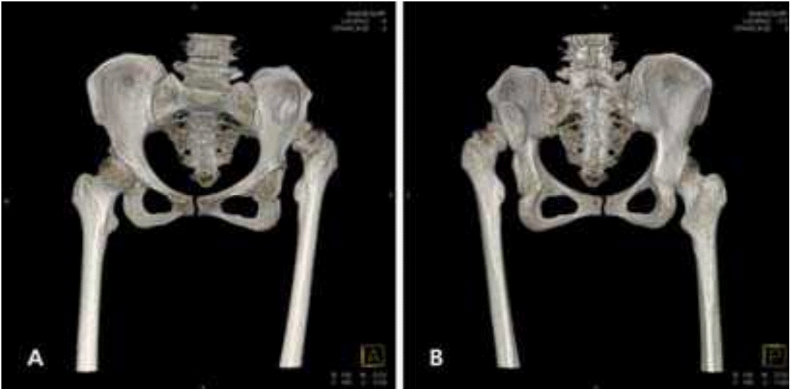
Computed tomography scan of the pelvis shows articulation of the left femoral head with the false acetabulum in the ilium more clearly. (A) Anteroposterior view (B) posteroanterior view.
Subtracting the femur from the CT scan, the false acetabulum in the ilium displayed a round and deepened articulating surface (Fig. 3.).
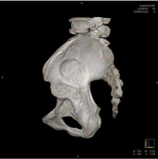
Left lateral view of a computed tomography scan of the pelvis bone shows the round and deepened false acetabulum in the ilium.
DISCUSSION
A shortened limb with adduction, internal rotation, and flexion of the hip joint is a typical presentation of posterior hip dislocation in trauma patients. However, developmental hip dislocation is a common complication in polio patients, and other various conditions can also cause developmental hip dislocation. If a patient has a history of limping gait since early childhood, congenital or developmental dysplasia of the hip should be suspected and careful radiologic evaluation is important to prevent misdiagnosis [1, 2].
Notes
CONFLICT OF INTEREST
No potential conflict of interest relevant to this article was reported.