Warning: mkdir(): Permission denied in /home/virtual/lib/view_data.php on line 86 Warning: fopen(/home/virtual/tipjournal/journal/upload/ip_log/ip_log_2024-04.txt): failed to open stream: No such file or directory in /home/virtual/lib/view_data.php on line 88 Warning: fwrite() expects parameter 1 to be resource, boolean given in /home/virtual/lib/view_data.php on line 89 Pneumocephalus caused by Compressed Air Injury without Fracture
TIP Search

CLOSE


Trauma Image and Procedure > Volume 2(1); 2017 > Article
Kim, Yun, and Gu: Pneumocephalus caused by Compressed Air Injury without Fracture

Abstract

Pneumocephalus and orbital emphysema without fracture or sinusitis is a rare condition. A 64-year old man suffered from right orbital trauma by a compressed-air-gun shooting. The right half of his face was swollen and a deep laceration of the right medial canthus was observed. Computed tomography showed extensive subcutaneous emphysema in the right periorbital soft tissue through the right temporal and zygomatic areas to the right upper neck, and disseminated pneumocephalus.

CASE

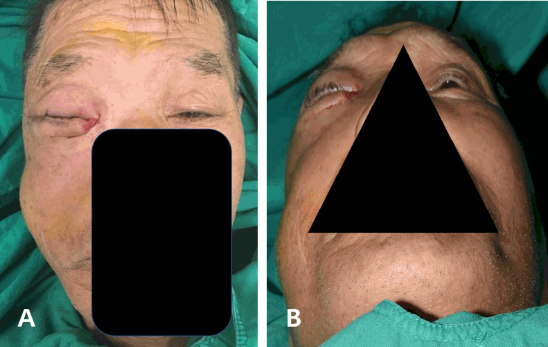
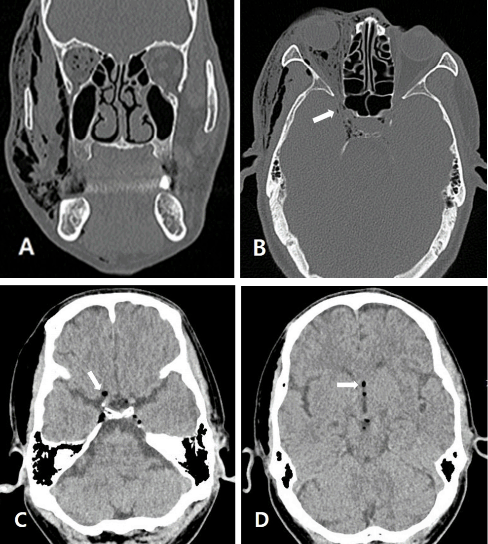
A 64-year-old man was admitted with swelling in the right periorbital area and right face and conjunctival hemorrhage with blurred vision in the right eye (Fig. 1), which had been exposed to a compressed air gun. Physical examination revealed conjunctival laceration, iritis, and corneal erosion in the right eye, a deep laceration of the right medial canthus, and right lacrimal canaliculi division. Computed tomography (CT) scan of the brain and facial bone showed extensive subcutaneous emphysema in the right periorbital area, which spread to the right temporal, retrobulbar, masseter, buccal, parapharyngeal, submandibular, and left periorbital areas, and disseminated pneumocephalus was noted in the subarachnoid space of the skull base without fracture of the facial bone and skull (Fig. 2.). There was no neurologic symptom or sign caused by the pneumocephalus. Lacrimal duct reconstruction for the right lacrimal canaliculus injury was performed on hospital day 2. A broadspcetrum antibiotic was applied for the pneumocephalus. Follow-up CT scan of the brain on hospital day 6 showed complete resolution of pneumocephalus and markedly decreased emphysema in the right face. The patient was discharged on hospital day 9 with improving blurred vision.

DISCUSSION

Although traumatic pneumocephalus with orbital emphysema usually is associated with skull fracture or paranasal sinusitis, compressed air injury often causes pneumocephalus without skull and facial bone fracture [1]. Emphysema caused by compressed air (high-pressure) exposure can spread extensively to various regions, including the orbit, face, neck, mediastinum, and intracranial cavity [2]. The mechanism of the air flow into the cranial cavity from the orbital cavity comes in through the dissection beneath the Tenon fascia, around the optic nerve, and through the optic canal into the subarachnoid space [3,4]. Treatment of the pneumocephalus is mostly via conservative therapy with systemic and topical prophylactic antibiotics. Prophylactic antibiotic therapy has an important role in preventing intracranial infection through the air entry route, because the protective barriers of the brain have broken down. The various emphysema and visual acuity conditions may resolve to normal within several days to 1 month [2].

Notes

Conflict of Interest Statement

No potential conflict of interest relevant to this article was reported.

Fig. 1.
An external photograph of the face reveals swelling of the right eyelids and cheek, and laceration of the right medial canthus (A, B).
tip-2-1-10f1.gif
Fig. 2.
CT scans of the face and brain. (A) Coronal CT of the facial bone shows the right orbital emphysema and extensive subcutaneous emphysema of the right cheek. (B) Axial CT of the facial bone shows pneumocephalus in the sella along the optic canal. (C, D) Brain CT shows disseminated pneumocephalus along the basal cistern (arrow) and third ventricle (arrow).
tip-2-1-10f2.gif

REFERENCES

1. Chung HJ, Lee BH, Hwang YJ, Kim SY. Pneumocephalus due to Compressed Air Injury without Facial Bone Fracture. Journal of the Korean Society of Radiology. 2011;65(4):333-5.
crossref
2. Hiraoka T, Ogami T, Okamoto F, Oshika T. Compressed air blast injury with palpebral, orbital, facial, cervical, and mediastinal emphysema through an eyelid laceration: a case report and review of literature. BMC ophthalmology. 2013;13(1):68.
crossref pmid pmc pdf
3. Yuksel M, Yuksel KZ, Ozdemir G, Ugur T. Bilateral orbital emphysema and pneumocephalus as a result of accidental compressed air exposure. Emergency radiology. 2007;13(4):195-8.
crossref pmid
4. Williams TR, Frankel N. Intracerebral air caused by conjunctival laceration with air hose. Archives of Ophthalmology. 1999;117(8):1090-1.
pmid
TOOLS
Share :
Facebook Twitter Linked In Google+
METRICS Graph View
  • 1 Crossref
  •    
  • 8,158 View
  • 54 Download


ABOUT
ARTICLE CATEGORY
Article Category

Browse all articles >

BROWSE ARTICLES
FOR AUTHORS AND REVIEWERS
Editorial Office
Korean Association for Research, Procedures and Education on Trauma
#1618, 18, Mapo-daero 4da-gil, Mapo-gu, Seoul 04177, Korea
Tel: +82-2-3280-9013    E-mail: karpet@karpet.or.kr

Copyright © 2024 by Korean Association for Research Procedures and Education on Trauma.

Developed in M2PI

Close layer
prev next