Warning: mkdir(): Permission denied in /home/virtual/lib/view_data.php on line 86 Warning: fopen(/home/virtual/tipjournal/journal/upload/ip_log/ip_log_2024-04.txt): failed to open stream: No such file or directory in /home/virtual/lib/view_data.php on line 88 Warning: fwrite() expects parameter 1 to be resource, boolean given in /home/virtual/lib/view_data.php on line 89 Sonographic Diagnosis of Biloma in Severe Traumatic Liver Injury
TIP Search

CLOSE


Trauma Image and Procedure > Volume 4(1); 2019 > Article
Park, Kang, Seo, and Moon: Sonographic Diagnosis of Biloma in Severe Traumatic Liver Injury

Abstract

Biloma is a common complication in patients with traumatic liver injury. Biloma is usually confirmed by abdominal computed tomography (CT), and the first detection using abdominal ultrasonography is rare. We report a case of a patient with American Association for the Surgery of Trauma (AAST) grade IV liver injury according to the Organ Injury Scale (OIS) score, who underwent transcatheter arterial embolization on the day of admission and percutaneous drainage after the diagnosis of biloma on ultrasonography on day 5 after admission. In this case, biloma was observed as a hypoechoic to anechoic fluid collection in the hepatic dome on ultrasonography.

CASE

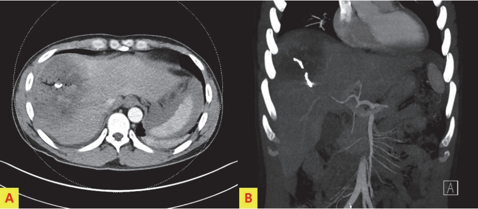
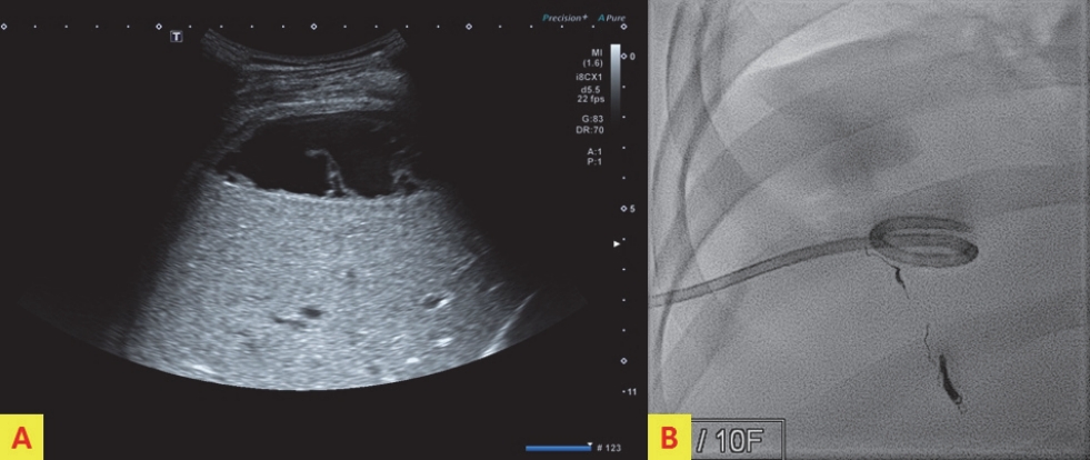
A 41-year-old man presented to the emergency department after a pedestrian traffic accident. The initial vital signs were unstable: systolic blood pressure, 80 mm Hg; pulse rate, 56 beats/min; respiration rate, 16 breaths/min; body temperature, 36.0℃; and oxygen saturation, 95%. Early abdominal computed tomography (CT) of the right side of the liver showed American Association for the Surgery of Trauma (AAST) grade IV injury, according to the Organ Injury Scale (OIS) score, with extravasation of contrast material (Fig. 1.). On the same day, hepatic artery angiography showed active bleeding in segments 5, 7, and 8, and embolization was performed (Fig. 2.). Liver enzyme levels (aspartate aminotransferase, 1036 IU/L; alanine transferase, 1192 IU/L) were highest on day 3 after admission. No extravasation of contrast material was observed on abdominal CT performed on day 3 (Fig. 3.). On day 5, the patient developed abdominal pain in the right upper quadrant. The focus of pain was more lateral than the location of the gallbladder on initial abdominal CT, and tenderness was also observed on physical examination. On abdominal ultrasonography, biloma was observed in the hepatic dome. Therefore, percutaneous drainage was performed (Fig. 4.). Biloma was still observed on abdominal CT 2 weeks later (Fig. 5.).

DISCUSSION

CT and ultrasonography play an important role in the initial treatment of abdominal blunt trauma. CT has high sensitivity for detecting liver parenchymal injuries. However, it is difficult to rule out organ injuries on ultrasonography, even when performed by skilled technicians [1]. Laboratory findings for biloma are nonspecific. If the right upper quadrant abdominal pain, chills, and fever are present in patients with recent abdominal injuries, biloma may be suspected [2]. Biloma appears as a hypodense fluid collection on CT [3]. Ultrasonography reveals a hypoechoic to anechoic fluid collection and well-defined margins findings in the typical location (right upper quadrant abdomen: sub-intrahepatic, below the diaphragm). Color Doppler ultrasonography shows no vascularity [4]. Garcia Urguelles et al. [5] stated that conservative management through percutaneous drainage under ultrasonographic guidance should be the first choice of treatment. In our patient, biloma was first diagnosed on ultrasonography after the patient reported right upper quadrant abdominal pain after blunt liver injury. Conservative management with percutaneous drainage was performed.

Notes

Conflict of Interest Statement

None of authors has a conflict of interest

Fig. 1.
Initial abdominal computed tomography, showing American Association for the Surgery of Trauma (AAST) grade II liver injury according to the Organ Injury Scale (OIS) score, with extravasation of contrast material in the right side of the liver.
tip-4-1-6f1.jpg
Fig. 2.
Hepatic artery angiography, showing active bleeding in segments 5, 7, and 8 (A). Embolization was performed at the bleeding focus (B).
tip-4-1-6f2.jpg
Fig. 3.
No extravasation of contrast material or biloma was observed on abdominal CT performed on day 3 after admission. The coils used for bleeding focus are visible.
tip-4-1-6f3.jpg
Fig. 4.
On day 5 after admission, biloma (hypoechnoic to anechoic fluid collection) is observed in the hepatic dome on ultrasonography (A). Percutaneous drainage was performed (B).
tip-4-1-6f4.jpg
Fig. 5.
Abdominal computed tomography 2 weeks after injury, showing biloma as a hypodense fluid collection. There is also drainage catheter in the biloma.
tip-4-1-6f5.jpg

REFERENCES

1. Poletti PA, Wintermark M, Schnyder P, Becker CD. Traumatic injuries: role of imaging in the management of the polytrauma victim (conservative expectation). Eur Radiol. 2002;12(5):969-78.
crossref pmid pdf
2. Tana C, D’Alessandro P, Tartaro A, Tana M, Mezzetti A, Schiavone C. Sonographic assessment of a suspected biloma: A case report and review of the literature. World J Radiol. 2013;5(5):220-5.
crossref pmid pmc
3. De Backer A, Fierens H, De Schepper A, Pelckmans P, Jorens PG, Vaneerdeweg W. Diagnosis and nonsurgical management of bile leak complicated by biloma after blunt liver injury: report of two cases. Eur Radiol. 1998;8(9):1619-22.
crossref pmid pdf
4. Colovic R, Perisic-Savic M. Retroperitoneal biloma secondary to operative common bile duct injury. HPB Surg. 1991;3(3):193-7.
crossref pmid pmc pdf
5. Garcia Urguelles X, Uroz Tristan J, Avila Suarez R, Alonso Jimenez L. Intrahepatic biloma. Conservative management with external drainage. Cir Pediatr. 1996;9(2):85-7.
pmid


ABOUT
ARTICLE CATEGORY
Article Category

Browse all articles >

BROWSE ARTICLES
FOR AUTHORS AND REVIEWERS
Editorial Office
Korean Association for Research, Procedures and Education on Trauma
#1618, 18, Mapo-daero 4da-gil, Mapo-gu, Seoul 04177, Korea
Tel: +82-2-3280-9013    E-mail: karpet@karpet.or.kr

Copyright © 2024 by Korean Association for Research Procedures and Education on Trauma.

Developed in M2PI

Close layer
prev next