Warning: mkdir(): Permission denied in /home/virtual/lib/view_data.php on line 86 Warning: fopen(/home/virtual/tipjournal/journal/upload/ip_log/ip_log_2024-04.txt): failed to open stream: No such file or directory in /home/virtual/lib/view_data.php on line 88 Warning: fwrite() expects parameter 1 to be resource, boolean given in /home/virtual/lib/view_data.php on line 89 Obstructive Atelectasis Caused by Total Obstruction of the Left Main Bronchus by Mucous Plug
TIP Search

CLOSE


Trauma Image and Procedure > Volume 3(2); 2018 > Article
Park and Kang: Obstructive Atelectasis Caused by Total Obstruction of the Left Main Bronchus by Mucous Plug

Abstract

Complete obstructive atelectasis caused by mucous plugs in the main bronchus can lead to life-threatening emergencies. Here, we report a case of atelectasis resulting in total obstruction of the left main bronchus due to a very large amount of thick purulent secretion, which was resolved using a fiberoptic bronchoscopic toilet.

CASE

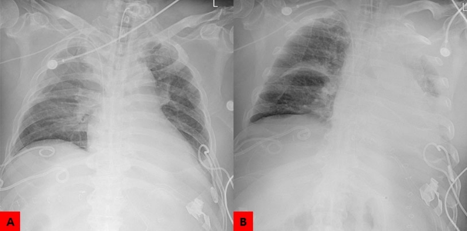
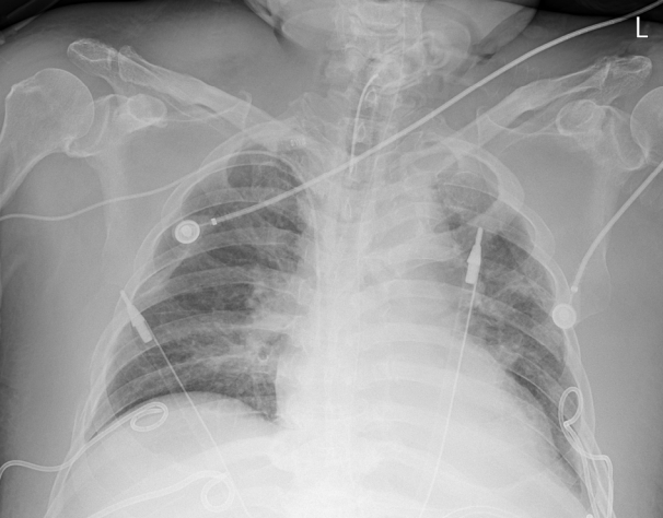
A 66-year-old male presented to the emergency department after suffering a major trauma. Endotracheal intubation and ventilator care were initiated on day 6 after hospitalization due to severe dyspnea. Open tracheostomy was performed on the 14th day of intubation. On the 20th day of tracheostomy, no atelectasis was observed in the AP chest radiography (Fig. 1A) and ABGA revealed the pH of 7.37, pCO2 of 36, pO2 of 121, HCO3 of 20.8, and O2 sat of 98.1. However, the total atelectasis in the left lung was observed in the AP chest radiography performed on the following day (Fig. 1B) and ABGA deteriorated to pH of 7.30, pCO2 of 42, pO2 of 54, HCO3 of 20.7, and O2 sat of 86.9. Fiberoptic bronchoscopy was performed to flush a very large amount of thick purulent secretion that totally obstructed the left main bronchus (Fig. 2). Atelectasis disappeared in the follow-up AP chest radiography (Fig. 3) and ABGA improved to pH of 7.32, pCO2 of 42, pO2 of 113, HCO3 of 21.6, and O2 sat of 99.2.

DISCUSSION

Atelectasis is a state in which the lungs are completely or partly collapsed, most commonly occurs postoperatively, and is caused by cystic fibrosis, lung tumors, chest injuries, lung fluids, respiratory weakness, and presence of foreign objects. Mucous plugs can also lead to atelectasis, and the main broncheal plug leads to complete pulmonary collapse, which can ultimately lead to life-threatening conditions [1]. Conventional chest radiography in a critically ill patient is the cornerstone of day-to-day management [2]. In addition, the possibility of atelectasis should be considered when O2 sat and pO2 changes occur. In this case, following a bronchoscopic toilet, O2 sat decreased from 98.1 to 86.9 and then increased to 99.2, whereas pO2 decreased from 121 to 54, and then increased to 113. Ghosh et al. [3] reported a case of left main bronchus obstructed by a chunk of thick mucous in a 6-year-old male, and a uniform opacity involving the whole lung field obscuring the heart shadow (Silhouette sign).

Notes

Conflict of Interest Statement

None of authors has a conflict of interest

Fig. 1.
On the 20th day of tracheostomy, no atelectasis was observed in the AP chest radiography. (A) However, total atelectasis in the left lung is observed in the AP chest radiography performed at the following day (B).
tip-3-2-56f1.jpg
Fig. 2.
Fiberoptic bronchoscopy reveals a very large amount of thick purulent secretion that totally obstructed the left main bronchus.
tip-3-2-56f2.jpg
Fig. 3.
Atelectasis in the left lung disappeared on the follow-up radiography following fiberoptic bronchoscopic toilet.
tip-3-2-56f3.jpg

REFERENCE

1. Coco D, Leanza S. Pleural Effusion or Main Left Bronchus Mucus Obstruction: To Drain or Not to Drain? Decision-Making for Young Surgeon on Call. Case Rep Radiol. 2018;2018:3180575.
crossref pmid pmc pdf
2. Khan AN, Al-Jahdali H, Al-Ghanem S, Gouda A. Reading chest radiographs in the critically ill (Part II): Radiography of lung pathologies common in the ICU patient. Ann Thorac Med. 2009;4(3):149-57.
crossref pmid pmc
3. Ghosh TR, Mandai MC, Das S, Mukhopadhyay MS, Basu SR. Left Main Bronchus Obstruction with Mucus Plug in a Child. J Anaesth Clin Phamacol. 2010;26(1):113-114.
crossref
TOOLS
Share :
Facebook Twitter Linked In Google+
METRICS Graph View
  • 0 Crossref
  •    
  • 9,790 View
  • 60 Download


ABOUT
ARTICLE CATEGORY
Article Category

Browse all articles >

BROWSE ARTICLES
FOR AUTHORS AND REVIEWERS
Editorial Office
Korean Association for Research, Procedures and Education on Trauma
#1618, 18, Mapo-daero 4da-gil, Mapo-gu, Seoul 04177, Korea
Tel: +82-2-3280-9013    E-mail: karpet@karpet.or.kr

Copyright © 2024 by Korean Association for Research Procedures and Education on Trauma.

Developed in M2PI

Close layer
prev next