Warning: mkdir(): Permission denied in /home/virtual/lib/view_data.php on line 86 Warning: fopen(/home/virtual/tipjournal/journal/upload/ip_log/ip_log_2024-04.txt): failed to open stream: No such file or directory in /home/virtual/lib/view_data.php on line 88 Warning: fwrite() expects parameter 1 to be resource, boolean given in /home/virtual/lib/view_data.php on line 89 Hepatic Hemangioma Mistaken As Intraparenchymal Hematoma of the Spleen
TIP Search

CLOSE


Trauma Image and Procedure > Volume 3(2); 2018 > Article
Ye, Go, Sul, Lee, Lee, Yoon, Kim, Lee, and Choi: Hepatic Hemangioma Mistaken As Intraparenchymal Hematoma of the Spleen

Abstract

Hepatic hemangioma is the most common benign liver tumor. Because most hepatic hemangiomas are asymptomatic, they are incidentally discovered during imaging investigations for other conditions. Although they were not found to be clinically meaningful, we report an interesting case of hepatic hemangioma, which was mistaken as intraparenchymal hematoma in the spleen because of its position and shape.

CASE

A 50-year-old male presented to the emergency department after meeting with a driver’s traffic accident. His mental status was alert, with all vital signs within normal ranges. The patient only complained of pain in the left lower chest wall.
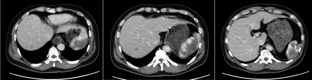
The findings of the focused assessment for sonography in trauma were all negative, and chest X-ray did not reveal any thoracic injuries. An abdominal computed tomography (CT) scan was subsequently performed, and it revealed an intraparenchymal hematoma in the spleen (Fig. 1).
The patient was hospitalized and closely monitored. The organ perceived to be the spleen was then found to be hepatic tissue extended from the left liver. In addition, the intraparenchymal hematoma was mistaken as hemangioma (Fig. 2).
In conclusion, hepatic hemangioma was mistaken as intraparenchymal hematoma in the spleen because of its position and shape. An atrophied spleen was observed between the tissue of hepatic hemangiomas, but the exact underlying cause is unknown (Fig. 2). The patient recovered with pain management and was discharged a few days later.

DISCUSSION

A typical hepatic hemangioma is detected as a hypodense, well-defined lesion on a CT scan, which shows peripheral nodular enhancement with progressive centripetal homogeneous filling after contrast injection [1]. Conversely, the typical intraparenchymal hematoma in the spleen can be seen on a CT scan as a low-density fluid adjacent to the spleen that distorts the splenic architecture [2]. Therefore, these two diagnoses can be easily distinguished by performing contrast-enhanced abdominal CT.
However, the location of hepatic hemangioma, the atrophied spleen, and the symptoms in this case may lead to the hemangioma being mistaken as intraparenchymal hematoma in the spleen. Thus, imaging findings should be more carefully examined to prevent unnecessary additional tests and long-term hospitalization.

Notes

Conflict of Interest Statement

None of authors have a conflict of interest.

Fig. 1.
An abdominal CT scan demonstrating spleen-like tissue with intraparenchymal hematoma in the left upper quadrant.
tip-3-2-47f1.jpg
Fig. 2.
Serial axial (A1–A5) and coronal views (B1–B4) of the CT scan show the hepatic hemangioma extended from the left liver in the left upper quadrant. An atrophic tissue in the spleen was also observed between hepatic hemangiomas (indicated by white arrows).
tip-3-2-47f2.jpg

REFERENCE

1. Bajenaru N, Balaban V, Savulescu F, Campeanu I, Patrascu T. Hepatic hemanioma. Journal of medicine and life. 2015;8:4-11.
pmid pmc
2. Hassan R, Abd Aziz A, Md Ralib AR. Computed tomography of blunt spleen injury: a pictorial review. Malays J Med Sci. 2012;18:60-7.

TOOLS
Share :
Facebook Twitter Linked In Google+
METRICS Graph View
  • 0 Crossref
  •    
  • 4,197 View
  • 35 Download


ABOUT
ARTICLE CATEGORY
Article Category

Browse all articles >

BROWSE ARTICLES
FOR AUTHORS AND REVIEWERS
Editorial Office
Korean Association for Research, Procedures and Education on Trauma
#1618, 18, Mapo-daero 4da-gil, Mapo-gu, Seoul 04177, Korea
Tel: +82-2-3280-9013    E-mail: karpet@karpet.or.kr

Copyright © 2024 by Korean Association for Research Procedures and Education on Trauma.

Developed in M2PI

Close layer
prev next